History ofLasik
Dr. Craig F. Beyer is an internationally recognized, board-certifed
ophthalmologist with extensive specialty training in laser refractive surgery.
As a member of the American Academy of Ophthalmology, the International Society for Refractive Surgery, the American Society of Cataract and Refractive Surgeons and the American Society for Laser Medicine and Surgery, Dr. Beyer brings years of experience, training and innovation to every surgery he performs.
Trained at the world-renowned LSU Eye Center, Dr. Beyer was a member of the surgical team that performed the first human excimer laser procedure in the world. He and his professor, Dr. Gholam Peyman, submitted the original paper for publication describing the LASIK procedure, which is now the most commonly performed surgical procedure in the world.
In 1990, Dr. Beyer became one of the first of 10 surgeons in the United States authorized by the FDA to perform investigational excimer laser surgery. He has performed laser vision correction longer than any other surgeon in the state of Colorado. Prior to excimer laser surgery approval in the United States, Dr. Beyer traveled the world lecturing and training physicians in laser refractive surgery. He is also the founder and former President of MedNet International, which manages eye surgery centers throughout China.
Similar to using crutches to walk after breaking a leg, approximately half of the population uses eyeglasses or contact lenses as a crutch to see. But, if you broke your leg, can you imagine how you would feel if your doctor told you that you would have to walk with crutches for the rest of your life? Well, for many years as a practicing ophthalmologist3, I told my patients that very same thing “I know you can’t see well without your glasses, but there’s nothing I can do – here are your glasses (crutches)”. Prior to laser vision correction, there weren’t many alternatives around for people who needed to wear glasses or contact lenses.
Those of us who don’t wear glasses or contacts can’t fully understand the miracle of LASIK surgery. People with good unaided vision take for granted the simple, daily tasks that are extremely cumbersome for eyeglass and contact lens wearers. To Put things into perspective, try to imagine not being able to see your clock every time you wake up from sleep to make sure you didn’t miss hearing your alarm. For men, ever tried lathering up and shaving your face with glasses on? Or, how would you feel if you had a contact lens dried onto your eye while snow skiing or driving down the highway on a motorcycle at 70 mph. These are just a few examples, the real hassles for eyeglass and contact lens wearers are much more numerous making their lives significantly more complicated than ours. Much of the free time we take for granted, others are busy searching for lost glasses or contacts, sterilizing lenses, buying cleaning solutions, following up with eye doctors, etc. Just so they can be prepared to take on the rest of the daily hassles in life.
In 1985, as a resident in ophthalmology, just like all other ophthalmology residents, I was trained to treat eye disorders such as cataracts4 and glaucoma5. But, what quickly became apparent to me was that we were not treating the most prevalent eye disorders of all – nearsightedness, farsightedness, and astigmatism (refractive errors) – these were the disorders the majority of my patients complained most about. Weren’t there any more permanent methods available to correct refractive errors? I knew about radial keratotomy8 (RK), but RK was not for everyone and only worked well for people with low degrees of nearsightedness. Ten, I reviewed an article written in the December 1983 issue of the American Journal of Ophthalmology by Steve Trokel, MD describing a new excimer laser that may someday be used to reshape the window to the eye (the cornea) to treat refractive errors. This was my first introduction to the idea of laser refractive surgery. Little did I know at the time that by 1988, I would be working directly with Steve and others to refine the laser that would perform the first human laser vision correction procedure in the world!

Dr. Steve Trokel (inventor of the excimer laser used for Laser Vision Correction) with Dr. Craig Beyer.
During the late 1970’s and early 1980’s, the excimer laser was used primarily by the computer industry to etch computer chips. The concept of using the excimer laser for an entirely different purpose, to treat biological tissue, was realized by two physicists independently, but nearly simultaneously.
During the late 1970’s, Dr. John Taboada was a physicist at Brooks Air Force Base in San Antonio. He was assigned by the military to research the safety and biological effects of using a new kind of laser called the excimer which generates invisible ultraviolet light in high energy pulses (the military was considering the use of ultraviolet light to communicate between satellites and submarines). In 1981, Dr. Taboada reported that the excimer laser created “craters” in experimental rabbit corneas. The craters took on the exact dimension and shape of the laser beam without harming the adjacent tissue. It is a matter of considerable controversy whether Dr. Taboada, at that time, realized the significance of his discovery related to laser vision correction (at issue, $80 million in past royalties).
Nearly at the same time, another physicist at IBM Research Center in New York, dr. R. Srinivasan, discovered the phenomenon of Ablative Photodecomposition11 (APD) using the excimer laser in 1908-81. His first report was entitled “Far-ultraviolet Laser Ablation12 of Atherosclerotic Lesions13. A second report entitled “Far-UV14 Photoetching of Organic material” was published in “Laser Focus” in May of 1983. To demonstrate APD in the most dramatic way, Srinivasan produced an electron micrograph of a highly magnified single human hair, in which precise cuts were etched by the excimer laser. Since people could relate to the heat sensitivity of hair, this image had a strong impact and was reproduced widely around the world. Each pulse from the laser removed a constant amount of tissue without any thermal damage to adjacent tissue without any thermal damage to adjacent tissue. Therefore, by simply counting the number of laser pulses, one could determine the amount of tissue removed within sub-micron accuracy. IBM was issued a patent in 1985 which broadly covered the use of the excimer on biological tissues but, crucially, didn’t mention eyes.
In 1982, Dr. Steve Trokel, an ophthalmologist who worked nearby IBM at Columbia Presbyterian Hospital was working with a different type of laser called the Nd:YAG laser. This laser remains in use today in cataract surgery. Hearing that Dr. Taboada had worked on the Nd:YAG laser, Trokel asked Taboada to write a chapter for a book. In March of 1983, Taboada met with Trokel and told Trokel about his excimer laser experiments on rabbit eyes. After learning about Taboada’s experiments, Trokel became interested in Srinivasan’s work at IBM. Trokel brought with him the Taboada articles and some veal eyes to test the effects of the excimer laser beam on the cornea.
Similar to Taboada’s work, the experiment revealed the ability to make precise incisions without harming adjacent tissue. Trokel was “awestruck” by the results and accurately foresaw that this would be the next great laser in ophthalmology. Within days, he flew to Europe to meet with the manufacturers of the IBM excimer laser to purchase his own laser.
Trokel was quick to file his own patent relating to refractive surgery and began refining his industrial laser for ophthalmic use. He formed a team of physicists, Charles Munnerlyn and Terry Clapham, to help him develop and commercialize this new concept. He consulted with two ophthalmologists Herb Kaufman, MD, Chairman of the LSU Eye Center in New Orleans and Marguerite McDonald, MD, also from LSU, who both earned leading international reputations in refractive surgery.

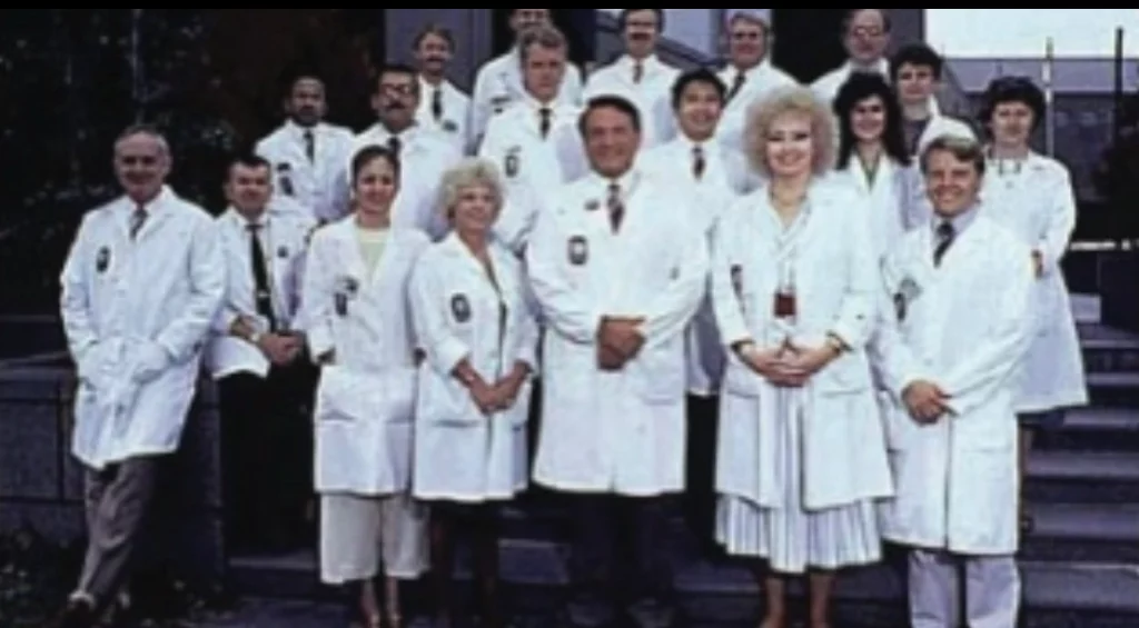
“The Team that Performed the First Laser Vision Correction Procedure in the World.” Drs Herbert Kaufman, Marguerite McDonald, and Craig Beyer.
In 1987, while presenting a paper at the Association for Research in Vision and Ophthalmology, I met Herb Kaufman who asked me “what would you think about a laser that in less than a minute could obviate the need for glasses or contacts forever?” Trying to cover-up the anxiety I felt after just speaking in front of hundreds of people and standing in front of one of the most respected ophthalmologists in the world, probably one of the simplest minded replies of all time slipped out of my mouth, “Wowwww!” Fortunately, I must have made a good impression upon Dr. Kaufman during my paper presentation, and just a few months later, he accepted me into his cornea and refractive surgery fellowship at the LSU Eye Center in New Orleans, LA; one of the most prestigious fellowships in the world.
In 1988, I began my refractive surgery fellowship at the LSU Eye Center in New Orleans, the same year Trokel assigned his patent rights to a new company called VISX, founded by Charles Munnerlyn. Also, this was the year VISX began testing its first prototype on humans at LSU. The very first patient to receive excimer laser surgery was a woman with uveal melanoma (a cancerous tumor that requires complete removal of the eye). Therefore, she voluntarily provided the opportunity for us to test the laser since there was little potential of doing additional harm. A week after the procedure, her eye was removed and studied histopathologically15 for any evidence of laser injury to the internal structures. Similar to Trokel’s and Taboada’s early experiments in animal eyes, no damage occurred to adjacent or internal tissues of the eye. Tus the Food and Drug Administration (FDA) allowed us to proceed with Phase 1 of the VISX investigational device exemption (IDE) on 10 blind eyes and then Phase II on 10 partially sighted eyes.
Oddly enough, unbeknownst to the unsuspecting ophthalmology resident, who examined her preoperatively, one of the blind patients was hysterically blind. In other words, there was no physiologic reason for her blindness. On her postoperative examination, after driving a car to the clinic, she pronounced she could see and proceeded to read 20/20 of the eye chart. Needless to say there were a lot of wide-open jaws in the clinic that day. Not only did the laser relieve this patient’s subconscious effort to suppress vision, but in addition, she was the first to experience the miracle of laser vision correction. Fortunately, the laser vision treatment corresponded to the high degree of her preexisting nearsightedness.
During all of the animal experiments and clinical trials from 1985 to 1993, all of the procedures were performed as PRK (photorefractive keratectomy). For PRK16, the laser is aimed directly at the surface of the cornea removing an area of tissue approximately 6 mm in diameter. One of my professors at LSU, Gholam Peyman, MD thought that this resulted in too much damage to the surface of the cornea leading to delayed healing, increased pain, and increased risk for scarring and infection. I also felt that there must be factors released by the healing corneal surface (the epithelium) which caused the underlying cornea (the stroma17) to scar. Therefore, Peyman and I began performing experiments in 1988 using a solid state erbium laser under a thin fap of corneal tissue which is how LASIK is performed today. By making a fap, the laser treatment is performed on the inside of the cornea allowing the surface of the eye to remain relatively undisturbed, therefore, there is virtually no pain and healing occurs within hours instead of days to weeks. In 1991, we published the first paper describing the experimental LASIK technique on primates*. At that time, the procedure was very difficult since I had to make the fap manually, thus I hardly suspected that other surgeons would rapidly master and perform this technique. However, after Luis Ruiz, MD perfected the automated microkeratome18 to make a corneal fap, LASIK has now become the most commonly performed refractive procedure in the world!
In 1989, we began the PRK studies on sighted eyes at LSU. As mentioned above, LASIK surgery was not even considered for the reason of increased surgical difficulty. The PRK results were exceptional for people with nearsightedness of 6.00 diopters19 or less. Above that number, PRK typically resulted in corneal scarring (today, with LASIK, we can successfully treat up to 12.00 diopters of nearsightedness). Encouraged by these early results, the FDA permitted VISX to open 10 additional experimental sites in the US where 75 patients could be treated at each site. In 1990, upon completion of my fellowship, I moved to St. Louis where I became one of the 10 VISX excimer laser principal investigators. I completed my portion of the study in 1992 and it wasn’t until 1995 that the FDA approved PRK for the treatment of nearsightedness.
From 1992 to 1995, I traveled to numerous countries training other surgeons in laser vision correction. Since other countries were not restricted by an entity similar to the FDA, PRK started to become popular with surgeons in South America, Europe, and Canada. However, it still did not seem to become too popular with patients. I had several patients who had undergone RK on one eye and then later, decided to have PRK on the other eye. I was somewhat astonished to learn that most of these patients preferred their RK eye! They said the pain was less, and their vision returned faster. I quickly realized that Dr. Peyman and I were right back in 1989. LASIK would solve these problems.
Therefore, in 1994, I flew to meet with Luis Ruiz who demonstrated to me the use of his new automated microkeratome. He was already performing hundreds of LASIK procedures each month and the results were astonishing. I quickly mastered his technique and became one of the early US surgeons to adopt the procedure and begin performing LASIK abroad (we still did not have FDA approval in the US for the excimer laser even for PRK). In China, where more than 70% of the population is nearsighted, I established 4 LASIK clinics where several hundred procedures are performed at each clinic every month.

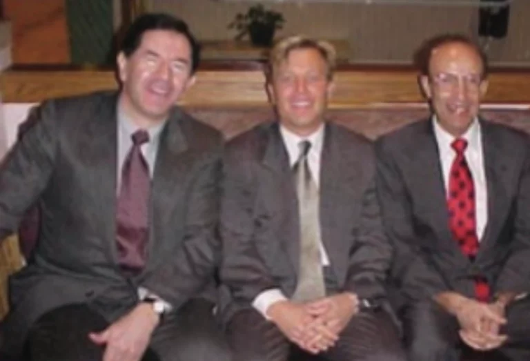
“The Founders of LASIK” (pictured from left to right: Dr Luis Ruiz, inventor of the microkeratome used to make the flap for LASIK, Dr Gholam Peyman, inventor of the LASIK procedure, and Dr. Craig Beyer, co-author and co-investigator (with Dr. Peyman) of the original scientific article describing the LASIK procedure.
After 2011, due to significant advances in PRK surgery, the recovery for PRK became much more acceptable and comfortable for patients. In addition, the use of Mitomycin C (that is applied at the time of PRK surgery) has reduced the risk for corneal haze (or scar tissue formation) to almost nil. Today, PRK actually carries less surgical risk and more long-term stability over LASIK; however, the downside is that PRK requires a slightly longer recovery than LASIK. All said, after 1-3 months, the results are essentially the same for both procedures. In our practice, we perform up to 40% of laser vision correction procedures as PRK since it is best suited for those with thin corneas, younger ages, low prescriptions, or for those who anticipate eye trauma such as professional boxers and law enforcement officers.